

Diabetes Dictionary
Diabetes is an endocrine disorder which is very complex and often takes time to fully understand. There is a great deal of information to learn as you begin to understand the terms and mechanics of diabetes.
Visit the American Diabetes Association Diabetes Dictionary for an easy-to-understand dictionary of common terms which are very useful to enhance your understanding of diabetes.
Pericytes are special cells that envelop the surface of the smallest blood vessels known as capillaries. Capillaries lie deep within the retina and serve as the conduit between arteries and veins. The role of the pericytes is one of maintenance and stabilization of the walls of the capillaries. Chronic elevated blood sugar levels (hyperglycemia) is believed to cause 'pericyte dropout'. The demise of pericytes has severe consequences because the walls of the capillaries weaken and generate microaneurysms. Research reveals that untrastructural analysis of retinal microaneurysms shows a consistent absence of pericytes, suggesting that the loss of capillary integrity due to the absence of pericytes likely renders capillaries venerable to microaneurysms leading to retinal disease and atrophy.
A microaneurysm is a weakened area of the capillary wall which gives way to an abnormal bulge or 'ballooning' in the wall of the capillaries. This ballooning of the capillary wall is liken to an old garden hose or worn out radiator hose. If we see a large bulge in the radiator hose, we know that it is just a matter of time before that weakened spot bursts. The eye doctor may not be able to visibly see these microaneurysms but they are very apparent upon a diagnostic test known as fluorescein angiography. An eye doctor is able to see bleeding if these microaneurysms give way and bleed into the retina.
Neovascularization of blood vessels is the formation of new blood vessels. The formation of new blood vessels in the retina is in response to a hypoxic or lack of oxygen situation which is related to capillaries collapsing due to disease of the walls of the capillaries. This disease process is triggered from too much sugar accumulating on the walls of the capillaries which starts a cascade of biologic events that leads to atrophy of the vessel and eventual closure.
This lack of oxygen is sensed by the retina. The retina responds by producing new blood vessels from areas of healthy retina attempting to reach oxygen-starved areas of retina. The problem with this valiant attempt by the retina is that these new blood vessels are only one cell thick and have no strength or stability. These new, fragile blood vessels are very vulnerable and may easily break or leak thereby spilling blood into the middle of the eye, the cavity known as the posterior chamber. The posterior chamber is normally filled with a clear fluid called vitreous. Should these new blood vessels break, a patient would notice very cloudy or darkened vision. The blood in the vitreous may take two or more months to clear or it may need to be evacuated.
Macular degeneration is the degeneration of retinal tissue in the area of retina known as the macula. This medical condition generally afflicts older adults and may result in the loss of central vision. This degenerative process occurs in a 'wet' or 'dry' form. The cause is still not known. There have recently been some new treatments and medications approved to treat macular degeneration. Research is ongoing into the cause and treatment.
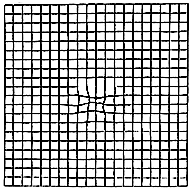
The Amsler Grid is a simple test to determine by the patient if there is a disturbance in the most vision sensitive area of the retina known as the macula. Swiss eye doctor Marc Amsler developed this grid in 1944 as he was studying the retina and doing pioneering work to map the field of vision. Amsler came to realize that any change in the macula tissue could be detected by the patient by viewing a grid on a chart. The test has the patient view a simple grid line chart, noting if there was any distortion of the grid lines or any waviness in the otherwise straight grid lines.
This chart has become the world standard in monitoring of the macula area. The grid is very much in wide use in eye doctors' offices today.

Macula area of retina.
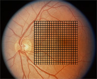
The grid area is seen as it is superimposed over the entire macula area.

The Amsler grid. Click here to go to the instruction
page and a printable version of the Amsler grid.
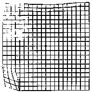
The Amsler grid as seen by the patient if there is a disturbance in the macula tissue.
Fluorescein angiography is a diagnostic technique that has been around since the 1960's and provides valuable information about the circulation system in the retina.
The test is performed by first injecting fluorescein dye into the vein in the arm. The dye takes just seconds to travel to the blood vessels of the retina. A camera equipped with a special filter to highlight the dye records high speed images as the dye courses through the circulation system of the retina. Images of the patterns of the dye will reveal if there is any swelling of the retina, leaking of the blood vessels or any other retinal abnormalities.
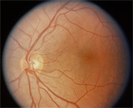
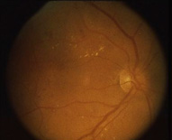
Normal retina as seen by the doctor.
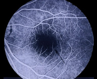
Normal retinal circulation as seen by fluorescein angiography.
Abnormal retina as seen by the doctor.
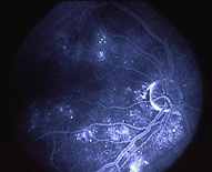
Abnormal retinal circulation system as seen highlighted by fluorescein dye. Note areas of leakage.
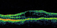
Optical Coherence Tomography (OCT) is a new imaging technique which produces a cross sectional image of the retina. The retina actually is composed of ten different layers of tissue, each layer having a specific function. Each retina is scanned with infra red light which is reflected back into the instrument. Each layer of retinal tissue has a specific reflectance value which is interpreted by the instrument and this reflectance is then translated into an image. The doctor then may view this cross section of retina to determine if any problem exists. The instrument takes a mere three seconds to scan each retina.
Normal OCT scan
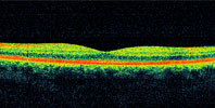 Here we can view the cross section of the retina. The different colored horizontal bands represent different layers of retinal tissue. The dip in the middle of the image is normal as the center of the dip represents the special area of the retina known as the macula. This is an image of a normal retina.
Abnormal OCT scan
 Here we have an image of an abnormal retina. This is an example of swelling in the macula area in the retina. Notice the raised area in the middle. This dark area represents fluid as fluid does reflect light and therefore presents dark. This area is also raised which indicates swelling of the tissue. The vision in this eye is poor with a very guarded, limited prognosis.